Human stem-cell laboratory model
Paper published 2017-04-26The biological question
Building a brain requires more than producing neurons. The cells must reach the right locations and join the right circuits. A Stanford-led study created a way to observe part of that process using living human cells grown in the laboratory.
Building a model of development
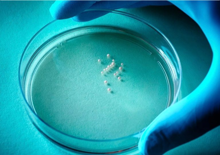
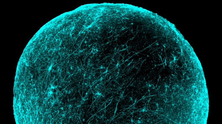
The researchers started with human pluripotent stem cells and guided them into three-dimensional clusters called spheroids. Different growth conditions produced tissue resembling two forebrain regions: a cortical region containing glutamatergic neurons and a ventral region containing GABAergic neurons. During development, GABAergic interneurons normally travel toward the cortex, where they become part of circuits that regulate neural activity.
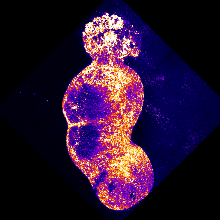
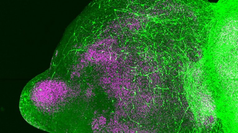
By bringing the two kinds of spheroids together, the team could watch that movement directly. The tissues fused, and interneurons crossed from the ventral-like spheroid into the cortical-like tissue. Live imaging revealed a stop-and-start pattern known as saltatory migration. The cells extended processes, moved forward, paused and repeated the sequence. Electrical recordings then showed that migrating neurons could integrate with the cortical neurons and participate in functional signaling.
Testing a disease mechanism
The study also used cells from people with Timothy syndrome, a rare genetic disorder associated with a calcium-channel mutation and neurodevelopmental differences. In the assembled tissues, the affected interneurons made abnormally frequent but less effective migratory movements. The work linked a genetic alteration to a measurable cellular behavior that is otherwise difficult to observe in the developing human brain.
Experiments that modified calcium-channel activity improved aspects of this migration phenotype in the dish. That result helped test the biological mechanism. It did not establish that these compounds treat Timothy syndrome in people, or that correcting one behavior in cultured neurons would correct every effect of the condition.
The platform's value lies in making a hidden developmental event experimentally accessible. Researchers can compare cells from different donors, change specific conditions and follow the consequences over time, rather than relying only on fixed tissue images.
What the model leaves out
There are clear boundaries to the model. Spheroids reproduce selected features of brain tissue; they do not contain the full organization, blood supply, sensory experience or developmental environment of a human brain. Different measurements also use different numbers of cells and donor lines, so a large cellular measurement is not equivalent to a large patient study. Within those limits, the paper offered a practical method for studying how developing human neurons migrate, connect and respond to genetic changes.
Key terms
- Spheroid
- A three-dimensional cell cluster modeling selected tissue features.
- Interneuron
- A neuron that participates in local circuits.
- Saltatory migration
- Cell movement in intermittent forward steps.
Original publication
Birey, F. et al. Assembly of functionally integrated human forebrain spheroids. Nature 545, 54–59 (2017).
Published online April 26, 2017.
Read the paper · DOI 10.1038/nature22330 ↗Stanford’s account of the study ↗
Sources reviewed & Stanford connection
Primary paper and official Stanford account reviewed.
Stanford-led research from Sergiu Pașca and collaborators.
This is an original explanatory article about the cited study. It is not a new paper or Stanford-authored news report.