Preclinical laboratory study
Paper published 2025-04-09The biological question
A sensory neuron does not deliver information directly to a fully formed experience. Signals pass through successive populations of cells, and the behavior of that relay can differ from the behavior of any neuron studied alone. A Stanford-led team asked whether the major components of the human ascending sensory pathway could be grown separately from pluripotent stem cells, connected in a dish, and studied together as a functioning network.
How the experiments worked
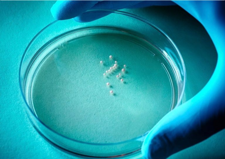
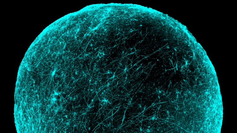
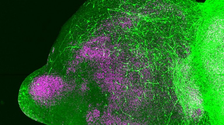
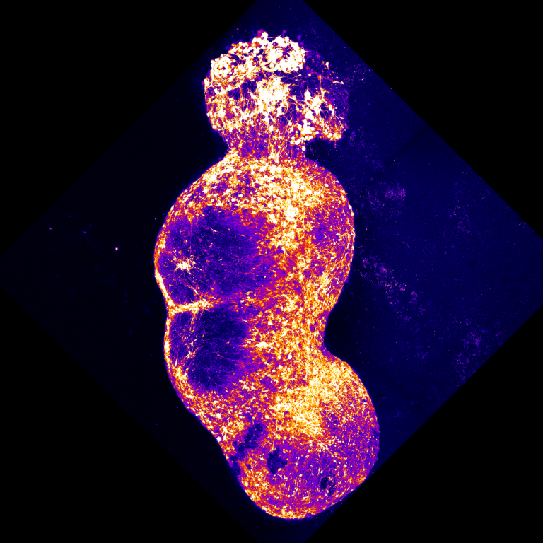
The researchers generated four regionalized organoid types: peripheral somatosensory tissue, dorsal spinal cord or hindbrain tissue, thalamic tissue, and cerebral cortical tissue. They used developmental signals to guide each population before bringing the parts together into a human ascending somatosensory assembloid. Single-cell RNA sequencing and protein staining tested whether the components contained cells with the molecular identities needed for the intended relay. Comparisons with developing human tissue supplied additional context for those identities.
Identity alone cannot establish that a circuit works. The team therefore combined anatomical labeling, modified-rabies tracing, optogenetic stimulation, calcium imaging, and electrical recordings. These approaches answer different questions: where projections extend, which cells form connections, whether stimulating one population can activate another, and whether activity becomes coordinated across the assembled system. The combination is stronger than treating a microscopy image of touching tissues as proof of functional communication.
What the researchers found
The assembled tissues formed connections along the modeled pathway. Chemical stimulation of sensory neurons could elicit coordinated activity across the construct, while recordings and imaging detected synchronized activity involving its constituent regions. These observations revealed an emergent property of assembly: the behavior of the relay became measurable across several linked human neural populations. The study also found a pharmacological difference between human sensory organoids and mouse sensory tissue in their response to the receptor antagonist TNP–ATP.
The investigators then altered SCN9A, the gene encoding the sodium channel NaV1.7. In people, certain loss-of-function variants cause profound pain insensitivity, while gain-of-function variants can cause severe pain syndromes. Removing functional NaV1.7 disrupted synchronization across the assembloid; a gain-of-function variant associated with extreme pain increased synchrony. The result moves the question beyond whether a mutation changes individual-cell excitability: it shows how that change can affect coordinated communication through a larger pathway.
Why the biology matters
This provides a biological reason to study circuits as assembled systems. A sensory neuron may still show activity without effectively recruiting downstream populations in the right temporal pattern. A candidate treatment might therefore need evaluation at both cellular and network levels. Because the model uses human cells, it also offers a way to investigate mechanisms that may differ across species. Those possibilities establish a research platform rather than demonstrating that a new pain treatment works in patients.
Limits and unanswered questions
The construct models an early developmental stage and a selected sensory pathway. Its sensory neurons have an incomplete repertoire of receptor expression, and the organoids do not recreate every neuronal type, anatomical arrangement, or feedback connection present in a person. The authors identify maturation and the specificity of connections as areas needing further work. The assembloid does not establish conscious pain: it lacks the broader systems required for the subjective experience and its emotional meaning.
The next useful questions are experimental. Can the sensory populations mature enough to represent later-life physiology? Which connections are consistently reproduced across donor cell lines and batches? Can a compound normalize a disease-associated network pattern without broadly suppressing neural activity? And do results in this simplified human pathway predict what happens in intact organisms? The study makes those questions accessible, while leaving therapeutic prediction and clinical benefit to future validation.
Key terms
- Assembloid
- A model formed by integrating separately generated three-dimensional tissues.
- NaV1.7
- A voltage-gated sodium channel encoded by SCN9A that contributes to sensory-neuron function.
- Synchrony
- Coordinated activity across cells or regions; it does not by itself imply conscious perception.
Original publication
Kim, J.-i., Imaizumi, K. et al. Human assembloid model of the ascending neural sensory pathway. Nature 642, 143–153 (2025).
Published online 2025-04-09.
Read the paper · DOI 10.1038/s41586-025-08808-3 ↗Read the peer-reviewed paper ↗
Sources reviewed & Stanford connection
Open Nature full text, main results and discussion read; official Stanford Medicine report used for affiliation and context.
This is an original explanatory article about the cited study. It is not a new paper or Stanford-authored news report.